qEEG Brain Mapping
Quantitative EEG — the brain map every protocol at Restorative Neuro is built on. Objective. Reproducible. Re-imaged at every milestone.
WHAT IT IS
What qEEG actually is.
qEEG (quantitative electroencephalography) records and analyzes the electrical activity of your brain at high temporal resolution. The recording is then compared to a normative database, producing topographic maps that show where and how your brain’s activity deviates from population norms. Unlike structural imaging (CT, MRI) which shows anatomy, qEEG shows function — how the brain is actually operating in time.
The technology has been clinically used for decades, and is a powerful diagnostic pillar in functional neurology.

WHAT A SESSION LOOKS LIKE
What a qEEG session looks like.
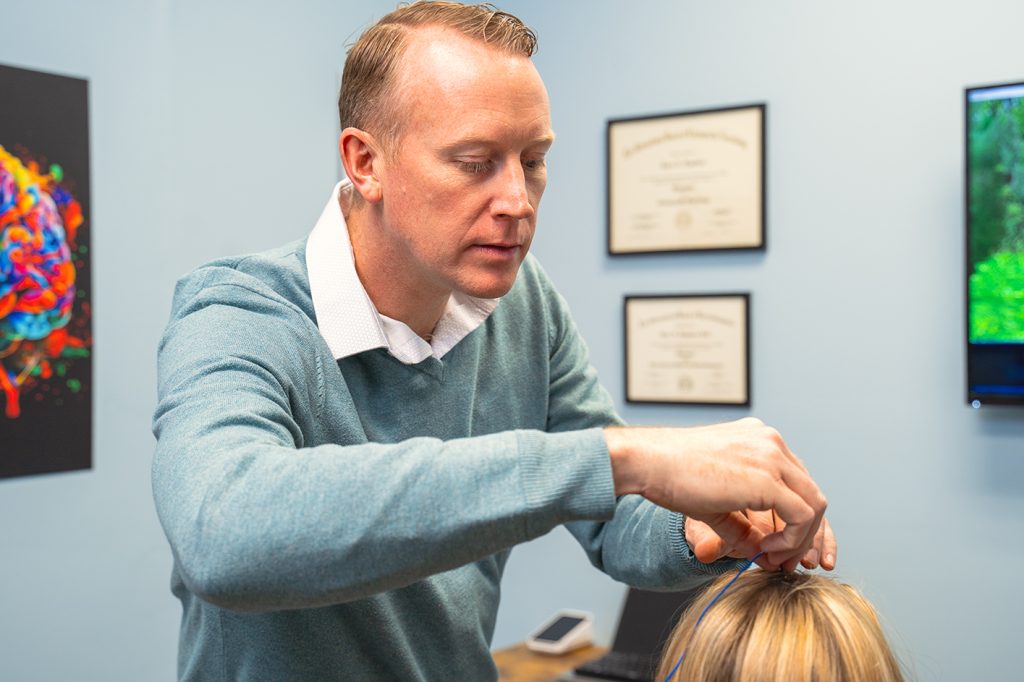
You sit in a comfortable chair. A cap with 19 sensors is placed on your scalp; a conductive gel is applied to each sensor (it washes out easily afterward). You sit quietly with eyes closed for several minutes, then with eyes open for several more. There is no electrical input — the sensors are recording, not stimulating. The whole recording takes about 30–45 minutes.
The analysis takes longer than the recording. Dr. Landers reviews the topographies, compares them to the normative database, and correlates findings with your symptom timeline and clinical exam. You receive a written report and an in-person review of the findings.
CONDITIONS WE USE THIS FOR
Indicated for the patterns above — selected from your brain map.
Post-Concussion Syndrome
qEEG surfaces network dysregulation MRI cannot show.
Brain Fog
Quantifies the cognitive-network signal.
ADHD
Identifies attention-regulation patterns.
EVIDENCE
Evidence base.
qEEG is supported by decades of peer-reviewed literature in neurorehabilitation, ADHD, mood disorders, and TBI. The research shows the effectiveness of brain maps and neurofeedback, and our modality stack ensures even greater functional gains.
Cost: for a complete qEEG including report and review. HSA/FSA eligible. Superbills available for out-of-network insurance reimbursement.
Research
-
Arns M, Clark CR, Trullinger M, et al. Neurofeedback and Attention-Deficit/Hyperactivity-Disorder (ADHD) in Children: Rating the Evidence and Proposed Guidelines. Applied Psychophysiology and Biofeedback. 2020;45(2):141-166. Concluded that standard neurofeedback protocols meet criteria for a “well-established” treatment for ADHD, with medium to large effect sizes and 32–47% remission rates sustained at 6–12 months.
-
Chen PY, Su IC, Shih CY, et al. Effects of Neurofeedback on Cognitive Function, Productive Activity, and Quality of Life in Patients With Traumatic Brain Injury: A Randomized Controlled Trial. Neurorehabilitation and Neural Repair. 2023;37(5). — The largest and most rigorous RCT to date (n=87), comparing neurofeedback to usual care. Neurofeedback significantly improved immediate recall, delayed recall, recognition memory, selective attention, and productive activity compared to controls.
-
Fernández-Alvarez J, Grassi M, Colombo D, et al. Efficacy of Bio- and Neurofeedback for Depression: A Meta-Analysis. Psychological Medicine. 2022;52(14):2801-2815. Found a large between-group effect size (Hedges’ g = 1.05) for neurofeedback in patients with major depressive disorder, with effects significant specifically in RCTs.
-
Annaheim C, Hug K, Stumm C, et al. Neurofeedback in Patients With Frontal Brain Lesions: A Randomized, Controlled Double-Blind Trial. Frontiers in Human Neuroscience. 2022;16:933506. — The first double-blind, sham-controlled RCT of neurofeedback in brain injury recovery (n=20). Showed significant improvement in the Frontal Assessment Battery and intrinsic alertness with active neurofeedback
-
Pindi P, Houenou J, Piguet C, Favre P. Real-Time fMRI Neurofeedback as a New Treatment for Psychiatric Disorders: A Meta-Analysis. Progress in Neuro-Psychopharmacology & Biological Psychiatry. 2022;119:110605. — Meta-analysis of 31 clinical trials of RT-fMRI neurofeedback across psychiatric disorders. Between-group analysis showed a large effect on anxiety (Hedges’ g = 0.77, p = 0.01).
-
Konicar L, Radev S, Prillinger K, et al. Volitional Modification of Brain Activity in Adolescents With Autism Spectrum Disorder: A Bayesian Analysis of Slow Cortical Potential Neurofeedback. NeuroImage: Clinical. 2020;29:102564. — RCT (n=41) of slow cortical potential (SCP) neurofeedback in male adolescents with ASD. The experimental group showed a 21.4-point reduction on the Social Responsiveness Scale (SRS). EEG analysis revealed a continuous decrease in delta power and increase in alpha power across training sessions, with non-linear trajectories suggesting complex neuroplastic changes.
-
Friedrich EV, Sivanathan A, Lim T, et al. An Effective Neurofeedback Intervention to Improve Social Interactions in Children With Autism Spectrum Disorder. Journal of Autism and Developmental Disorders. 2015;45(12):4084-4100. — Pre-post study (n=13) of 16 sessions of mu-rhythm neurofeedback using a social interaction game paradigm. Children showed improvements in mu suppression, emotion recognition, spontaneous imitation, and everyday behavior, demonstrating that neurofeedback paradigms targeting social cognition can improve components necessary for successful social interactions.